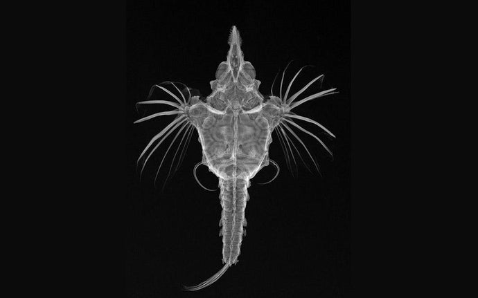
Fish Radiography

The Artistry of Fish Radiography
At times, science is an artform unto itself. So it is for a series of x-rays of fish by the Smithsonian Maritime Museum that have allowed scientists to build a comprehensive narrative of the diversity of fish. Tracing the changes in number of vertebrae, fin position and countless other variations, the striking sequence dives through to the depths of fish evolution. Compiled by the Smithsonian Institute in Ichthyo - The Architecture Of Fish, it is a stunning compendium of science and art.
The Scientific Method
X-ray images allow the study of fish anatomy without dissection or alteration of the specimen. Radiographs may be prepared for any number of any species, so allowing a researcher to compare various features both intra- and inter-species. Images are designed to follow scientific convention, not artistic flair. There is always one specimen per frame, and the fish is always facing to the left. A beam of x-rays is generated and focused on the specimen. As tissue density determines absorption of x-rays, the bones of the specimen materialise most readily, creating the images. Characteristics of the species can then be readily seen, assessed and compared. Relative size and shape of bones and fins, the presence or absence of teeth, biological elements are tabulated and archived.
Art From Science, Philosophy From Art
From these images, the beauty, intricacy and diversity of the patterns of nature become self-evident. It is a natural union between science and art. The metaphysical element of evolution emerges from the arranged sequence, the sublime artistry of constant adaptation through mutation. One abstracts to the general nature of biology - of the continuous change, of the possibilities of the future, the direct line of ancestors - it is a sombre thought. One realises their position in the great chain of life, as an infinitesimal yet influential piece of the greatest whole.


Words | Rob Woodgate


